How do chromosomes divide?
In a body, every single cell has the same number of chromosomes. This is because when a cell divides, the replicated chromatids are equally segregated into daughter cells. But, the first meiotic division is a clear exception. This division segregates maternal and paternal chromosomes for production of eggs and sperms, which are the origin of a new life. What is the story behind this division?
From a single cell
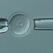
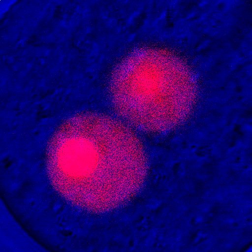
Your body initially began as a single cell. This cell, called a zygote, was generated by the fusion of an egg with a sperm. The egg and sperm were produced in your mother and father, respectively. The egg was derived from an oocyte, which had been stored in the ovary of the mother.
Oocyte
The oocyte is a specialized cell. The oocyte becomes an egg through meiosis, a special type of cell division, and serves as a starting point for embryonic development once fertilization takes place. To enable these mysterious and dramatic processes, the oocyte has specific features that are not seen in other cell types.
How chromosomes segregate
Among the many events that take place in oocytes, we are particularly interested in chromosome segregation, one of the most fundamental cellular events. Chromosomes carry genetic information. Oocytes first undergo meiosis I, in which maternal and paternal chromosomes are segregated. The oocytes then undergo meiosis II upon fertilization, during which chromosome segregation occurs with no DNA replication. Subsequently, the fertilized egg (zygote) undergoes the first cell division, mitosis, in which replicated chromosomes are segregated. We aim to understand the mechanisms underlying these three successive, but fundamentally different, chromosome segregation events. Through this study, we will be able to understand the drama that unfolds in the oocytes - how strategies and machinery for chromosome segregation are prepared, properly used, and at times fail .
Errors in chromosome segregation
Oocytes are, in fact, not good at segregating chromosomes in meiosis I. Errors are estimated to occur in 10-30% of chromosome segregation during meiosis I in oocytes. The eggs produced with errors have an abnormal number of chromosomes, which results in pregnancy loss or, if survives to term, results in congenital diseases such as Down syndrome.
Errors increase with age
The frequency of segregation errors in oocytes increases with maternal age. It is estimated that, in clinically recognized pregnancies, the frequency of trisomy in embryos is approximately 5% at 30 years old, 8% at 35 years old, and 25% at 40 years old (Hassold & Hunt 2001 Nat Rev Genet). In most of these cases, the trisomy originates from errors in meiosis I.
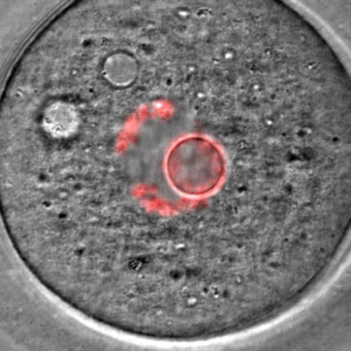
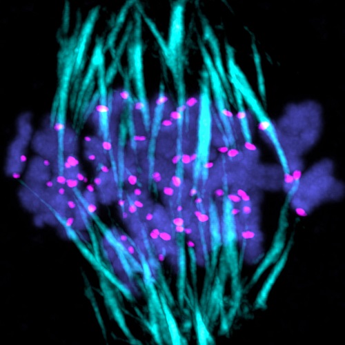
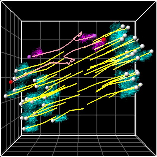
Filming chromosome segregation
Team leader Tomo Kitajima succeeded in carrying out a detailed analysis of the chromosome dynamics during meiosis I in mouse oocytes when he worked as a postdoc in the laboratory of Dr. Jan Ellenberg at EMBL Heidelberg (Kitajima et al 2011 Cell). He captured live images of chromosome dynamics in living oocytes using automated microscopy at high resolution.
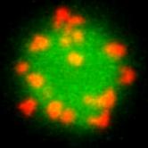
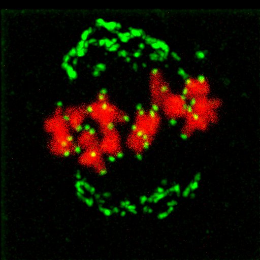
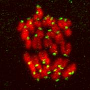
Chromosome belt
Four-dimensional analysis of oocytes revealed a belt-like spatial arrangement of the chromosomes prior to formation of stable attachments between chromosomes and microtubules. This arrangement might help chromosomes to dictate the microtubules to attach to proper spindle poles.
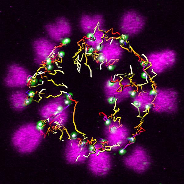
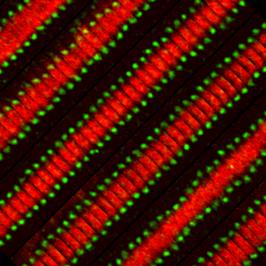
Error-prone attachment
After forming the chromosome belt, microtubules start attaching to chromosomes to pull them to the spindle poles. However, microtubules frequently form incorrect chromosome-microtubule attachments, and correct attachments are not easily stabilized. Our team recently proposed that this meiosis I-specific chromosome property contributes to the error-prone nature of chromosome-microtubule attachment (Yoshida et al 2015 Dev Cell).
Correct attachments
To achieve proper chromosome segregation, chromosome-microtubule attachments must be corrected. How can these attachments be corrected? Further investigation on this issue js needed, but so far we know at least of one requirement to correct errors; that is tension generated across chromosomes when microtubules pull the chromosomes to opposite poles. The tension somehow eliminates errors and stabilizes correct attachments. However, oocytes do not appear to have difficulties to setting this requirement.
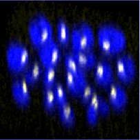
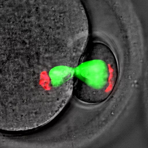
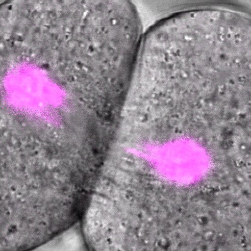
Age-related errors
With advanced maternal age, some chromosomes become incapable of generating proper tension. Our team recently succeeded in directly observing single chromosomes undergoing segregation errors in living aged oocytes (Sakakibara et al 2015 Nat Commun). In young oocytes, chromosomes can generate tension when pulled to opposite spindle poles by microtubules. In aged oocytes, however, when microtubules pull the chromosomes to opposite poles, the chromosomes are unable to resist the opposing forces and prematurely separate into two pieces. These separated pieces have no way to generate proper tension, and thus, result in chromosome segregation errors.
And next?
We plan to reveal the mechanisms underlying chromosome segregation during the process of oocytes becoming a fertilized eggs, taking advantage of established high-resolution live imaging of mouse oocytes combined with micromanipulation and genetic engineering techniques. Our studies provide a basis for the development of clinical approaches to infertility and congenital diseases.
![]()
![]()